


A Guide to Clinical Differential Diagnosis of Oral Mucosal Lesions
Course Number: 110
Course Contents
Part II: Surface Lesions of Oral Mucosa
Remember that surface lesions of oral mucosa consist of lesions that involve the epithelium and/or superficial connective tissue. They do not exceed 2-3 mm in thickness. Clinically, surface lesions are flat or slightly thickened rather than being swellings or enlargements.
We initially divide surface lesions into three categories based on their clinical appearance: white, pigmented, and vesicular-ulcerated-erythematous.
White Surface Lesions of Oral Mucosa
Surface lesions of oral mucosa that appear white, tan, or light yellow are divided into three groups based on their clinical features:
White lesions due to epithelial thickening
White lesions due to accumulation of necrotic debris on the mucosal surface
White lesions due to subepithelial changes in the connective tissue.
Epithelial thickening white lesions appear white because the pink to red color of the blood vessels in the underlying connective tissue is masked by the increased thickness of the epithelium. These lesions are asymptomatic, rough to palpation, and cannot be rubbed off with a gauze. They appear flat white when dried.
Three of the epithelial thickening white lesions occur on the tongue: hairy tongue, hairy leukoplakia, and geographic tongue (erythema migrans).
Hairy tongue* is the result of the accumulation of keratin on the dorsal surface of the tongue. Numerous causes have been proposed, but lack of mechanical stimulation to the dorsal tongue due to poor oral hygiene and/or a soft diet are probably the most important causes. The lesion presents as elongated filiform papillae having a hair-like appearance. The papillae are typically stained brown, black or other colors depending on the patient’s diet and habits. Hairy tongue is typically not painful. Hairy tongue is not a serious condition, but warrants treatment for cosmetic and hygienic reasons. Treatment involves using a toothbrush, tongue blade, or tongue scraper to brush or scrape the dorsal surface of the tongue. The prognosis is good.
Hairy tongue
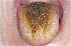
Hairy tongue
Hairy leukoplakia is caused by Epstein-Barr virus and presents as unilateral or bilateral, asymptomatic, white, rough patches, usually on the lateral surfaces of the tongue. It most commonly occurs in HIV positive patients but can also be found in any immunocompromised patient. Hairy leukoplakia does not require treatment, but it should alert the clinician that the patient is immunocompromised.
To view the Decision Tree for Oral Mucosal Lesions, click on one of the options shown.
 View Interactive
 View as PDF
 View as GIF
To view the Decision Tree for Oral Mucosal Lesions, click on one of the options shown.