


A Guide to Clinical Differential Diagnosis of Oral Mucosal Lesions
Course Number: 110
Course Contents
Idiopathic Diseases
Idiopathic diseases are those of unknown or poorly understood cause. These diseases do not have common historical and clinical features as a class or group, and thus each disease must be considered individually in the differential diagnosis.
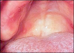
Aphthous ulcers* are a common cause of recurrent oral discomfort. The ulcers have an abrupt onset and resolve in a predictable amount of time for each patient, usually 7-14 days. Aphthous ulcers occur on nonkeratinized mucosal surfaces, such as buccal and labial mucosa, ventral surface of the tongue, floor of the mouth, and soft palate. A familial history is sometimes reported. The ulcers may be menstrually related. “Major” aphthous ulcers are larger and of longer duration than typical aphthae and often heal with scarring. “Herpetiform” aphthae refer to multiple crops of small aphthous ulcers.
Aphthous ulcers
Aphthous ulcers are most commonly treated with topical corticosteroids, such as triamcinolone acetonide, 0.1% mouthrinse. A short burst of systemic corticosteroids may be needed for persistent lesions. Intralesional injection of corticosteroids can also be of value for a larger, deeper lesion. It is important to explain to the patient that the goal of therapy is to control the lesions, and that there is currently no definitive cure.
Erosive lichen planus* is a chronic inflammatory disease involving skin and oral mucosa. It represents an immune abnormality involving T lymphocytes directed against antigens in the overlying stratified squamous epithelium. Occasionally lichen planus is associated with medications the patient is taking. The skin lesions consist of pruritic (itching), erythematous to light purple patches, sometimes with white striations. Oral lesions most commonly appear as white epithelial thickening arranged in a network pattern (Wickham striae) with erythema of the surrounding mucosa. White patches, erythematous erosions, and ulcers may occur with, or in place of, the striae. The white lesions are asymptomatic, but the erosions and ulcers are usually painful. Lichen planus almost always has multiple lesions bilaterally, with the buccal mucosa commonly involved. Oral lesions may occur without skin lesions. Asymptomatic lesions require no treatment other than inspection during annual dental visits. Topical and/or systemic corticosteroids will almost always control, but not cure, painful erosions and ulcers of lichen planus. If suspected lichen planus is refractory to traditional treatment, an incisional biopsy may be required for definitive diagnosis.
Erythema multiforme* is an idiopathic disease that involves an immunologic abnormality. It may be triggered by infection, especially with herpes simplex virus, or drugs, such as antibiotics. It is characterized by the acute onset of blisters and ulcers of skin and oral mucosa. The appearance of the skin lesions is variable. “target” or “iris” lesions of the skin are characteristic but not present in all cases, and consist of a blister surrounded by erythematous rings. Oral mucosal blisters and ulcers are present in multiple locations and are painful. Hemorrhagic crusting of the lips is often present. Fever, malaise, and pharyngitis may precede the lesions.
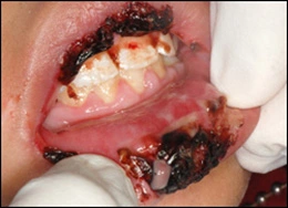
Erythema multiforme
Stevens-Johnson syndrome is a severe form of erythema multiforme and demonstrates conjunctivitis and genital ulcers in addition to mucocutaneous lesions. Topical and/or systemic corticosteroids may be useful in treatment. Offending drugs should be discontinued. If it appears to be associated with herpes simplex infection, then anti-viral medications may be used. Erythema multiforme is typically benign and resolves spontaneously, although it may recur. Rarely, Stevens-Johnson syndrome may be fatal.
Medication-induced mucositis*: A number of different medications cause oral mucosal lesions that do not appear to be allergic reactions but rather represent a toxic side-effect of the medication. These mucosal lesions can present as nonspecific ulcers, erosions or may resemble erosive lichen planus. They occur on both keratinized and nonkeratinized mucosal surfaces, and are chronic lesions. They do not necessarily appear immediately after the patient begins taking the medication.
The first step in management is diagnosis. An incisional biopsy usually shows a nonspecific ulcer but may be useful in excluding other causes of chronic mucositis, such as erosive lichen planus and mucous membrane pemphigoid. After the diagnosis is made, the dentist should consult with the patient’s physician to see if a different medication can be used. Once the offending medication is withdrawn, the lesions resolve.
Medication-induced mucositis
Contact stomatitis: Numerous chemical agents in food, candy, chewing gum, toothpaste, and mouthrinses can cause chronic mucositis. Flavoring agents, especially cinnamon flavoring, are common causes of contact stomatitis. The clinical features of contact stomatitis from cinnamon flavoring include mucosal pain, burning, erythema, edema, erosion, and ulceration. Hyperkeratosis often covers the erythematous areas producing a shaggy thickened surface. The lesions are diagnosed based on the clinical features and a history of cinnamon exposure. The lesions typically resolve within one week following discontinuation of the cinnamon products. Rarely, amalgam restoration or metal materials in crowns can cause a lichen planus-like reaction in the adjacent mucosa.
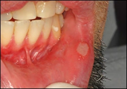
Erythroplasia (erythroplakia) is a clinical term corresponding microscopically to epithelial dysplasia, carcinoma in situ*, or superficially invasive squamous cell carcinoma*. It appears clinically as asymptomatic, persistent, erythematous, velvety, focal to diffuse mucosal areas. Because these lesions are asymptomatic, the patient is almost never aware of them.
To view the Decision Tree for Oral Mucosal Lesions, click on one of the options shown.
 View Interactive
 View as PDF
 View as GIF
To view the Decision Tree for Oral Mucosal Lesions, click on one of the options shown.