


A Guide to Clinical Differential Diagnosis of Oral Mucosal Lesions
Course Number: 110
Course Contents
Melanocytic Lesions
Melanocytic lesions appear brown or black due to the deposition of melanin.
Ephelis* is a freckle. It is flat, brown or black in color, and occurs on sun-exposed surfaces. It is due to increased production of melanin by melanocytes. An ephelis requires no treatment.
Oral melanotic macule* is a localized pigmented lesion associated with increased melanin pigmentation of the stratified squamous epithelium. It is asymptomatic, flat and not thickened, and appears similar to an ephelis (freckle) of skin. It is a harmless lesion, but its significance lies in distinguishing it from nevus or early melanoma. A biopsy should be performed if any doubt exists about the diagnosis.
If an oral pigmented lesion is not thickened, but is larger in diameter, has any variation in color, cannot be diagnosed as tattoo based on radiographic findings, or has irregular borders it should be excised.
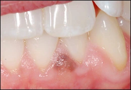
Oral melanotic macule
Melanocytic Nevus* is a benign proliferation of nevus cells (melanocytes). Nevi of skin first appear in childhood and progress through a series of clinical and microscopic stages. Most people have between 10 and 40 nevi on their skin. Nevi of skin that have uniform color and borders and are not changing in size or surface texture are not considered premalignant lesions and do not need to be removed unless they are chronically irritated or are a cosmetic problem. Nevi of oral mucosa are relatively rare. They occur most commonly on the gingiva and hard palate. Nevi of oral mucosa should be completely excised because they cannot be differentiated from melanoma based on their clinical features.
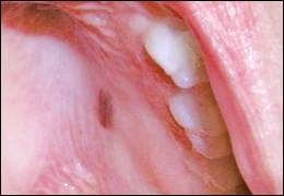
Nevus
Melanoma* is a malignant neoplasm of melanocytes. Melanoma of skin has increased significantly in incidence, while melanoma of oral mucosa is relatively rare. The most important clinical features of cutaneous melanoma are asymmetry of the lesion, variation in color (brown, black, red, white, blue), and diameter greater than 6 mm. Oral melanoma begins as an irregular, brown to black macule. Later the lesion will develop thickening and sometimes ulceration. The most common locations are the hard palate, gingiva, and alveolar ridge. It is not possible to distinguish an oral melanocytic nevus from early melanoma.
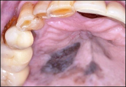
Melanoma
If oral nevus and/or melanoma are included in the clinical differential diagnosis, then a biopsy is indicated. Biopsy is also indicated for flat, non-thickened pigmentations that are changing or have atypical color, borders, or size. Treatment for melanoma is complete surgical excision. The thickness of the lesion and depth of invasion are the most important prognostic factors.
To view the Decision Tree for Oral Mucosal Lesions, click on one of the options shown.
 View Interactive
 View as PDF
 View as GIF
To view the Decision Tree for Oral Mucosal Lesions, click on one of the options shown.