


A Guide to Clinical Differential Diagnosis of Oral Mucosal Lesions
Course Number: 110
Course Contents
Pigmented Surface Lesions of Oral Mucosa
Pigmented surface lesions of oral mucosa appear blue, brown, or black. They are classified as generalized lesions, which are diffuse and multifocal, and localized lesions, which are unilateral and involve only one or several locations. Note that some soft tissue enlargements are pigmented, but they are discussed under Soft Tissue Enlargements.
Generalized Pigmented Surface Lesions of Oral Mucosa are bilateral, multiple and diffuse. There are numerous causes for generalized pigmentations, varying from common to rare, and the most important are discussed below.
Hereditary (racial, ethnic, physiologic)* pigmentation is the most common type of generalized pigmentation. The pigmentation is diffuse, symmetrical, and most apparent on the gingiva and labial mucosa. In general, the extent of oral mucosal pigmentation is directly related to the extent of skin pigmentation.
Pregnancy can lead to multiple melanotic macules on oral mucosa and facial skin (melasma or chloasma). No treatment is necessary for the melanin pigmentation, and it typically fades after the pregnancy.
Numerous medications*, such as quinine drugs used in the treatment of systemic lupus erythematosus, can cause diffuse pigmented lesions.
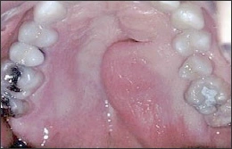
Smoker’s melanosis is caused by stimulation of melanin production by melanocytes due to chemical substances in cigarette smoke. The anterior facial gingiva is most commonly involved, although any oral mucosal site can demonstrate this. Often smoker’s melanosis can be clinically diagnosed by correlating a history of smoking with the location and distribution of the pigmentation. If the diagnosis is not evident, then biopsy is indicated. No therapy other than smoking cessation is necessary once the diagnosis has been made.
Ingestion of, or exposure to, heavy metals*, such as lead, mercury, gold, arsenic and bismuth, can lead to diffuse pigmentation of oral mucosa. The pigmentation may be dark blue, gray or black and commonly involves the marginal gingiva. Diffuse mucosal ulceration and a metallic taste may also be noted. Extraoral manifestations may be a clue to the diagnosis, and include dermatitis, tremors, mental changes, headache, fatigue, and gastrointestinal upset. Management of suspected heavy metal intoxication involves referral for diagnostic workup.
Peutz-Jeghers syndrome is a genetic condition characterized by numerous freckle-like lesions on the skin of the hands and around the mouth, nose and anogenital region. Intra-oral freckles may involve the lips, tongue and buccal mucosa. Patients also have multiple polyps, mainly in the small intestine. The polyps sometimes result in intestinal obstruction. Patients have an increased risk of gastrointestinal carcinoma but the polyps are not premalignant. Newly diagnosed patients with this syndrome should be referred for evaluation of the gastrointestinal tract
Neurofibromatosis (von Recklinghausen disease of skin) is a genetic disease with multiple subtypes. Type I is the most common type and is characterized by multiple neurofibromas. The neurofibromas vary in size, number, and may be well circumscribed or diffuse. Melanotic macules called café-au-lait spots, at least 1.5 cm in diameter and numbering 6 or more, are diagnostic of neurofibromatosis. Axillary freckles are also common. Numerous other systemic manifestations may be present in neurofibromatosis. Central nervous system abnormalities are especially prominent. There is no definitive treatment for neurofibromatosis. See also the discussion of neurofibromatosis with neurofibroma.
Neurofibromatosis
Polyostotic fibrous dysplasia is a systemic syndrome in which diffuse bony lesions of fibrous dysplasia involve multiple areas of the skeleton. The McCune-Albright syndrome includes polyostotic fibrous dysplasia, café-au-lait melanotic macules, and endocrine abnormalities, such as precocious puberty in females. The Jaffe-Lichtenstein syndrome includes polyostotic fibrous dysplasia plus café-au-lait pigmentation without endocrine abnormalities.
Note that the vast majority of cases of fibrous dysplasia of the jaws occur as a solitary (monostotic) lesion rather than as part of the polyostotic syndrome. Monostotic fibrous dysplasia does not have generalized café-au-lait pigmentations.
In Addison disease* the adrenal cortex is destroyed, resulting in decreased production of cortisol, aldosterone and adrenal androgens. Signs and symptoms include weakness, anorexia, nausea, vomiting, diarrhea, abdominal pain, decreased serum sodium, and hypotension. Diffuse pigmentation of skin and oral mucosa typically occur in Addison disease. Treatment consists of replacement therapy with glucocorticoids and mineral corticoids. The prognosis is good with appropriate therapy.
To view the Decision Tree for Oral Mucosal Lesions, click on one of the options shown.
 View Interactive
 View as PDF
 View as GIF
To view the Decision Tree for Oral Mucosal Lesions, click on one of the options shown.